

Cartilage Conditions
Expert Cartilage Treatment at the World's #1 Hospital for Orthopaedics in New York City
Articular cartilage injuries in the knee, hip, and shoulder, and osteochondritis dissecans — evaluated for cartilage-preserving treatment before joint damage progresses.
Cartilage Conditions
{{ c.anatomy }}
{{ c.condition }}
{{ c.classifications }}
{{ c.riskFactors }}
{{ c.symptoms }}
{{ c.diagnosis }}
{{ c.nonSurgical }}
{{ c.surgical }}
Cartilage Restoration of the Knee
Cartilage restoration treats a focal cartilage defect — a localized area of damage — with the goal of rebuilding the joint surface, relieving pain, and delaying or preventing progression to arthritis. Defects arise from sports injuries and dislocations, from developmental conditions such as osteochondritis dissecans, or from gradual wear.
There is no single best procedure. The right technique depends on the size, depth, and location of the defect, whether the underlying bone is involved, and the patient’s activity goals. Patients with widespread, bone-on-bone arthritis are usually better served by other treatment options.
What determines the right procedure
Techniques
Comparing the techniques
The choice of procedure depends on where the defect sits, how large it is, its shape, whether the bone underneath is involved, and what recovery a patient is able to take on. Not every procedure produces the same repair tissue. Marrow stimulation forms fibrocartilage, which is mechanically weaker than native cartilage; graft-based techniques restore tissue that behaves much more like the knee’s own surface. That distinction is why larger defects and those involving bone usually call for a graft.
| Treatment | Where it works best | Defect size | Defect and joint features | Bone involvement | Other considerations |
|---|---|---|---|---|---|
| Debridement | Any location in the joint | Any size | Best for irregular, elongated, or unstable flaps of cartilage, with or without joint swelling. Can be used when both sides of the joint are affected. | If the bone beneath is contributing to symptoms, it can be treated at the same time. | Suited to patients who want a shorter recovery, accept that relief may be partial, and place lower physical demands on the joint. |
| Marrow stimulation (with or without biologic augmentation) | Any location, though less commonly used on the kneecap | Small to medium defects | For contained defects that are unlikely to improve with debridement alone. Preferred when only one side of the joint is involved. | Underlying bone can be addressed in the same procedure if needed. | A reasonable option for defects whose shape or location makes other techniques less suitable. |
| Osteochondral autograft (OAT) | Femoral condyles, with attention to matching the joint surface shape; kneecap pain is a consideration when treating that region | Small; medium defects can be treated with multiple plugs when donor tissue is limited | For contained, round or oval defects that can be treated with minimal disruption of the surrounding healthy cartilage and bone. Preferred when one side of the joint is involved, but tolerates mild changes on both. | Restores both cartilage and the bone beneath it. | Often the preferred choice for small defects of the femur compared with marrow stimulation. |
| Osteochondral allograft (OCA) | Any joint surface other than the tibia | Medium to larger defects, where smaller ones have less resource-intensive options | For contained, partially contained, or complex-shaped defects. Preferred when one side of the joint is involved, but tolerates mild changes on both. | Well suited to defects with bone loss; large amounts of missing bone may be grafted in the same or a staged procedure. | Often preferred for revision cases, defects involving bone, and when combined with procedures such as meniscal transplantation. |
| MACI (cell-based cartilage implantation) | Any joint surface other than the tibia | Medium to large defects, where smaller ones have less resource-intensive options | A primary option for defects on one side of the joint, contained or partially contained, in multiple locations and of varying size — especially in younger patients and around the kneecap. | Best suited to defects with minimal change in the bone beneath the cartilage. | Depending on surgeon experience, often favored over allograft for kneecap-region defects, where the surface shape is complex. |
| Coral-based scaffold | Femoral condyles and trochlea (the groove at the front of the knee) | Small to larger defects | For contained defects, including elongated or irregular shapes that are less suited to other options. Tolerates mild changes on both sides of the joint. | Avoided when there is meaningful change or loss of the underlying bone. | May be considered in knees with more advanced arthritic change when other options are limited, though results are more variable. |
| Allogeneic articular cartilage surface graft | Any location | Medium defects, or larger ones when alternatives exist for smaller defects | A primary option for defects on one side of the joint, contained or partially contained, in multiple locations and of varying size — especially in younger patients and around the kneecap. | Avoided when there is meaningful change or loss of the underlying bone. | Insurance coverage can be limited, so use and published results are less extensive. |
Published work on cartilage
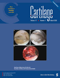
Dr. Kunze has authored extensive peer-reviewed and award-winning articles on cartilage injuries and treatments that is shaping our understanding of cartilage and joint preservation. His work has contributed to how physicians around the world treat these complex injuries.






Risks and benefits
- Relief of mechanical pain, catching, and swelling.
- Restoration of the joint surface rather than replacement of it.
- Preservation of the native knee in younger and active patients.
- In most cases, a realistic return to sport and demanding activity.
- General surgical risks: infection, blood clots, and anesthetic risk.
- Knee stiffness, or arthrofibrosis.
- Incomplete integration of the graft.
- Failure of the repair tissue to fill the defect.
- Progression of arthritis over time.
- Marrow-stimulation techniques form fibrocartilage, which can break down in larger or high-demand defects.
No cartilage procedure is a guaranteed permanent fix. These operations are designed to relieve pain, restore the joint surface, and delay or avoid joint replacement. Results depend on defect size and location, alignment, body weight, activity level, and adherence to rehabilitation — all of which are reviewed individually before a plan is made.
Follow-up and long-term outcomes
Follow-up is structured around the biology of the repair rather than the calendar. Early visits confirm motion is returning and that weight-bearing is progressing safely; later visits — typically at three, six, and twelve months — track function, strength, and readiness for higher-impact activity. Imaging is repeated when the clinical picture calls for it, usually to confirm graft integration before clearance for sport.
Over the longer term, graft-based restoration of well-selected focal defects has a durable track record: most patients report meaningful, sustained improvement in pain and function, and many remain active for years without progressing to joint replacement. Marrow-stimulation techniques tend to perform well early and less reliably over time in larger defects, which is why defect size and depth drive the choice of procedure in the first place.
Durability also depends on the rest of the knee. Correcting alignment, restoring meniscal function, and stabilizing ligaments at the time of surgery meaningfully improves how long a cartilage repair lasts. Patients are followed with standardized outcome measures so results can be compared against published benchmarks and used to guide future recommendations.
Disclaimer: All materials presented on this website are the opinions of Dr. Kyle Kunze and any guest writers, and should not be construed as medical advice. Each patient’s specific condition is different, and a comprehensive medical assessment requires a full medical history, physical exam, and review of diagnostic imaging. If you would like to seek the opinion of Dr. Kyle Kunze for your specific case, we recommend contacting our office to make an appointment.